Overview
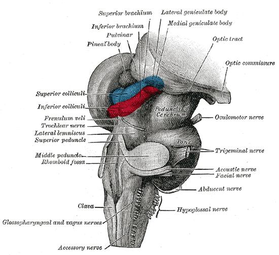
1.Lateralgeniculatenucleus:Thelateralgeniculatenucleusisanucleusoftheposteriorthalamus.Itislocatedontheventralsurfaceofthethalamus,theoutersideofthecerebralfeetandtheupperpartofthemedialgeniculatebody.Itconsistsoftwoparts,thelargerdorsalnucleusandthesmallerventralnucleus.Itistherelaycoreofthevisionsystem.Receivefibersfromtheretinaandemitfiberstoprojecttotheprimaryvisualcortex.
2.Diencephalon:Diencephalonislocatedbetweenthemidbrainandthetelencephalon.Thediencephalonandthetelencephalonbothderivefromtheforebrainalarintheearlyembryonicstage.Thediencephalonislocatedinthebackcenteroftheforebrain,andthetelencephalondevelopsintotheleftandrightcerebralhemispheres.Duetothehighexpansionofthetelencephalon,thediencephalonissurroundedbytheleftandrightcerebralhemispheresexceptforthepartthatbelongstothehypothalamusontheventralside,whichisexposedonthesurfaceofthebrain.Inthemid-sagittalsectionofthebrain,thelinefromtheposteriorcommissuretotheposterioredgeofthepapillarybodyrepresentsthejunctionbetweenthediencephalonandthemidbrain,andthelinefromtheinterventricularforamentotheopticchiasmrepresentsthejunctionbetweenthediencephalonandthetelencephalon.Theventricularcavityofthediencephaloniscalledthethirdventricle.

Theinnersideofthediencephalononbothsidesformsthesidewallofthethirdventricle.Atthejunctionofthemedialanddorsalside,thereisaraisedfiberbundle—thalamicmedullastriatum,whichisattachedtothethirdventriclechoroidtissue.Themedullaryveinsofthethalamusconnectthereintriangleback,andthereisareincommissureconnectionbetweentheleftandrightreintriangles,andthereisapinealglandbehindthiscommissure.Approximatelyinthemiddleofthethirdventriclewall,thereisanadhesive(orcentralmass)betweenthethalamusconnectingtheleftandrightventricularwalls.Ithasahypothalamicsulcusonitsventralside,fromthemidbrainaqueducttotheinterventricularforamen.Thestructurebelongingtothehypothalamussurroundsthefloorofthethirdventricle,andfromfronttobacktherearetheopticchiasm,thefunnel,thegraynodulesandthenipplebodyconnectingtheendplates.Onthebackofthediencephalon,bothsidesofthethirdventricleareheldbyovalgraymattermassesbelongingtothedorsalthalamus,withaprotrudinganteriorthalamicnoduleatthefrontendandanenlargedoccipitalattheback.Thereisthecaudatenucleusbelongingtothetelencephalonontheoutersideoftheback,andthestriaterminalisbetweenitandthediencephalon.Onthelowerandoutersideofthepillow,therearemedialgeniculatebodyandlateralgeniculatebody.Thelateralsurfaceofthediencephalonisfusedwiththeinternalcapsuleofthetelencephalon.Theventralsurfaceofthediencephalonisthepartexposedontheoutersurfaceofthebrain,withtheopticchiasmandoptictractbeforeit.Thefunnel,pituitaryandgraynodulesareinthemiddle,andthepapillarybodiesarepairedandlocatedbehindthegraynodules.
Anatomy
Lateralgeniculatebody:Thelateralgeniculatebodyislocatedunderthethalamusocciput,andthemedialgeniculatebodyisontheoutside,oneontheleftandoneontheleftandoneontheleftandoneontheleftandright.Thegroup,calledthelateralgeniculatenucleus,isthesubcorticalcenterofvision,connectedtothesuperiorcolliculusofthemidbrainquadrantviathesuperiorcolliculusarm.Thelateralgeniculatebodyisapairofcranialnervesdirectlyconnectedwiththediencephalon,thatis,theterminalnucleusoftheopticnerve(Ⅱ).Thefibersemittedbythenucleusformopticradiationandreachthevisualcenterofthecerebralcortex.Itisthelastrelaynucleusonthevisualconductionpathway..
Themorphologicalstructureandcompositionofthelateralgeniculatebody
Thelateralgeniculatebody:Thelateralgeniculatebodyisalsocalledtheexternalgeniculatebody.Thesmallellipticalbumpslocatedontheoutsideofthefeetofthebrainandundertheoccipitaloftheopticthalamusbelongtothediencephalonandarethefirst-levelvisualcenterofthevisualanalyzer.Theopticfibersoftheoptictractstopattheganglioncellsofthelateralgeniculatebodyandexchangeneuronstoformopticradiation,whichallprojecttothestriateareaofthevisualcenteronthesamesidetoproducevision.Theopticalfibersinthelateralgeniculatebodyalsohaveacertainarrangement.Theperipheraluncrossedfibersfromtheipsilateralretinaandthecontralateralcrossedfibersterminateontheventralsideofthelateralgeniculatebody.Theupperhalfofthefiberislocatedontheventralinnersideandthelowerhalf.Thefibersarelocatedontheventralside.Themacularfiberisfinallydorsal.Theupperquadrantmacularfiberislocatedinsidethewedge-shapedarea,andthelowerquadrantmacularfiberislocatedoutsidethewedge-shapedarea.VonMonakowdividesthelateralgeniculatebodyintothreeparts,namely:①thelowerpartoftheanteriorpart,whichisconnectedtotheopticbeam;②thedoorpart;③thelateralpart,whichisthespurofthelateralgeniculatebody.
Thecharacteristicsandclinicalsignificanceofthebloodsupplyofthelateralgeniculatebody
Thesmallarteriesofthelateralgeniculatebodyaremulti-sourceandmulti-vesselbloodsupply,80%ofwhicharemorethandualsources,smallarteriesAsmanyas3-6,thesesmallarteriesfromdifferentsourcesareanastomosedwitheachotheronthesurfaceofthelateralgeniculatebody.Webelievethatevenifthereare1-2tertiarybloodsupplyarteriesblockedornarrowed,itwillnotaffectthenutritionofthelateralgeniculatebodyandvisualfielddefects.Thepossibilityisunlikely.However,20%ofthesinglesourcearterialblooddonorshaveimportantsignificanceintheetiologyofvisualfielddefects.Forexample,thesourcearterialatherosclerosiscanblocktheartery,whichcancausethelateralgeniculatebodytobecomeischemicandlosetheconductionfunction.
